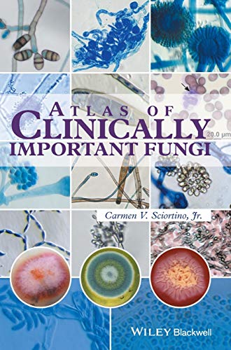

Atlas of clinically important Fungi
Detalles de publicación: Wiley-Blackwell 2017ISBN:- 9781119069669
Descripciones mejoradas de Syndetics:
Although there are many texts that provide quality information for the identification of fungi, researchers and technologists rarely have time to read the text. Most are rushed for time and seek morphological information that helps guide them to the identification of fungi.
The Atlas of Clinically Important Fungi provides readers with an alphabetical list of fungi as well as listing the division of fungi by both sporulation and morphology. The characteristic traits for a particular fungus are displayed through a series of images, with the fungi appearing as they did in the author's lab on the day(s) that testing was performed. For this reason, numerous (6-20) color photographs are included so that technologists will have sufficient reference photos for identifying the various morphologies of a single organism. Organism photographs begin with the macroscopic colony views followed by the microscopic views. Also included for some microorganisms, are clinical pathology photographs demonstrating how the organism appears in human tissues.
A collection of literature citations are also provided to enable further reading.
This user-friendly fungi atlas provides a resource for those seeking information in the field of medical mycology, specifically with regards to identifying an organism using the parameters of culture morphology.
Tabla de contenidos provista por Syndetics
- Preface(p. xi)
- Acknowledgements(p. xiii)
- Abbreviations(p. xv)
- Part I Divisions of the Fungi(p. 1)
- 1 Morphological Growth Index(p. 3)
- 1.1 Filamentous Bacteria(p. 3)
- 1.2 Yeasts and Yeast-like Organisms(p. 3)
- 1.3 Yeast/Yeast-like Organisms That Produce Hyphae(p. 4)
- 1.4 Thermally Dimorphic Fungi(p. 4)
- 1.5 Thermally Monomorphic Molds(p. 4)
- 1.6 Dematiaceous (Brown or Black Colored Fungi)(p. 6)
- 1.7 Other Fungi for Which No Photographs are Available(p. 8)
- 2 Division of Fungi by Type of Sporulation(p. 9)
- 2.1 Annelloconidia(p. 9)
- 2.2 Arthroconidia(p. 9)
- 2.3 Arthroconidia and Blastoconidia(p. 9)
- 2.4 Arthroconidia; Spherule in Tissue(p. 9)
- 2.5 Basidiospores(p. 9)
- 2.6 Phiaioconidia(p. 9)
- 2.7 Produce Only Macroconidia(p. 10)
- 2.8 Macroconidia and Microconidia(p. 10)
- 2.9 Perithecium and Ascospores(p. 10)
- 2.10 Sporangia or Sporangiospores(p. 10)
- 2.11 Sporangia with Rhizoids(p. 11)
- 2.12 Produce Only Microconidia(p. 11)
- 2.13 Microconidia and Chlamydoconidia(p. 11)
- 2.14 Microconidia and Arthroconidia(p. 11)
- 2.15 Microconidia and Cleistothecium(p. 11)
- 2.16 Yeast Only(p. 12)
- 2.17 Yeast and Hyphae and Phialides(p. 12)
- 2.18 Yeast and Pseudohyphae(p. 12)
- 2.19 Yeast and Conidia(p. 12)
- 3 Division of Fungi, Yeast, and Bacteria by Morphological Group(p. 13)
- 3.1 Actinomycetes, Yeast, and Yeast-like Organisms(p. 13)
- 3.2 Dimorphic Organisms(p. 13)
- 3.3 Dermatophytes(p. 13)
- 3.4 Zygomycetes (Aseptate)(p. 14)
- 3.5 Hyaline Molds (Septate)(p. 14)
- 3.6 Dematiaceous Fungi (Septate)(p. 14)
- Part II Descriptions and Photographs of Microorganisms(p. 17)
- 4 Actinomycetes, Yeast (Thermally Monomorphic), and Yeast-like Organisms(p. 19)
- 4.1 Microorganisms with Fungal-like Morphology Occurring in Fungal Cultures(p. 19)
- 4.2 Yeast and Yeast-like Fungi(p. 19)
- 4.3 Candida spp(p. 34)
- 4.4 Prototheca spp(p. 36)
- 4.5 Arthrographis kalrae(p. 39)
- 4.6 Cryptococcns neoformans(p. 44)
- 4.7 Geotrichum candidum(p. 47)
- 4.8 Malassezia spp(p. 56)
- 4.9 Trichosporon spp(p. 58)
- 5 Yeast (Thermally Dimorphic)(p. 63)
- 5.1 Blastomyces dermatitidis(p. 63)
- 5.2 Coccidioides immitis(p. 69)
- 5.3 Histoplasma capsulatum(p. 73)
- 5.4 Sporothrix schenckii(p. 83)
- 6 Zygomycetes (Aseptate)(p. 89)
- 6.1 Lictheimia (Absidia) corymbifera(p. 89)
- 6.2 Conidiobolus lamprauges, coronatus, incongruus(p. 95)
- 6.3 Cunninghamella bertholletiae(p. 102)
- 6.4 Mwcor spp(p. 106)
- 6.5 Rhizomucor spp(p. 116)
- 6.6 Rhizopus spp(p. 121)
- 6.7 Syncephalastrum racemosum(p. 130)
- 7 Dermatophytes(p. 135)
- 7.1 Microsporum spp(p. 135)
- 7.2 Trichophyton spp(p. 145)
- 7.3 Epidermophyton floccosum(p. 161)
- 8 Hyaline Molds (Septate)(p. 167)
- 8.1 Aspergillus spp(p. 167)
- 8.2 Beauveria bassiana(p. 185)
- 8.3 Emmonsia spp(p. 189)
- 8.4 Fusarium spp(p. 195)
- 8.5 Gliocladium(p. 217)
- 8.6 Malbranchea spp(p. 222)
- 8.7 Paecilomyces spp(p. 225)
- 8.8 Penicillium spp(p. 230)
- 8.9 Ramichloridium spp(p. 234)
- 8.10 Schizophyllum commune(p. 240)
- 8.11 Scopulariopsis brevicaulis(p. 246)
- 8.12 Scopulariopsis brumptii(p. 252)
- 8.13 Sepedonium spp(p. 256)
- 8.14 Sporotrichum pruinosum(p. 260)
- 8.15 Trichoderma spp(p. 266)
- 8.16 Trichothecium roseum(p. 273)
- 8.17 Chrysosporium tropicum(p. 276)
- 9 Dematiaceous Fungi (Septate)(p. 283)
- 9.1 Acremonium spp(p. 283)
- 9.2 Alternaria spp(p. 291)
- 9.3 Aureobasidium pullulans(p. 294)
- 9.4 Bipolaris spicifera, Curvularia hawaiiensis (B. hawaiensis), and C. australiensis (B. australiensis)(p. 299)
- 9.5 Chaetomium spp(p. 307)
- 9.6 Cladophialophora (Xylohypha) bantiana(p. 310)
- 9.7 Cladophialophora boppi(p. 313)
- 9.8 Cladophialophora carrionii(p. 316)
- 9.9 Cladosporium sphaerospermum(p. 319)
- 9.10 Cladosporium spp(p. 322)
- 9.11 Curvularia spp(p. 329)
- 9.12 Epicoccum spp(p. 334)
- 9.13 Exserohilum spp(p. 339)
- 9.14 Fonsecaea(p. 345)
- 9.15 Hormonema dematioides(p. 353)
- 9.16 Hortaea (Phaeoannellomyces, Exophiala) werneckii(p. 357)
- 9.17 Madurella spp(p. 363)
- 9.18 Neoscytalidium spp(p. 371)
- 9.19 Nigrospora spp(p. 378)
- 9.20 Ochroconis (Dactylaria) spp(p. 382)
- 9.21 Phaeoacremonium parasiticum(p. 387)
- 9.22 Phialemonium spp(p. 392)
- 9.23 Phialophora spp(p. 397)
- 9.24 Pithomyces sp(p. 403)
- 9.25 Pseudallescheria boydii(p. 409)
- 9.26 Scedosporium(p. 414)
- 9.27 Stachybotrys chartarum(p. 424)
- 9.28 Stemphylium sp(p. 428)
- 9.29 Ulocladium sp(p. 431)
- 9.30 Phoma sp(p. 435)
- 9.31 Rhinocladiella basitona(p. 441)
- 9.32 Exophiala dermatltidis (Wangella dermatitidis)(p. 447)
- 9.33 Exophiala jeanselmei(p. 453)
- Additional Reading(p. 459)
- Index(p. 467)