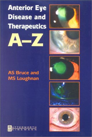

Anterior eye disease and therapeutics A-Z Adrian S. Bruce, Michael S. Loughnan.
Detalles de publicación: Oxford New York Butterworth-Heinemann 2003.Descripción: x, 358 p. ill. (chiefly col.) 24 cmISBN:- 0750652616
- 617.7 B8863a 21
- 2002 O-120
- WW 210
| Imagen de cubierta | Tipo de ítem | Biblioteca actual | Biblioteca de origen | Colección | Ubicación en estantería | Signatura topográfica | Materiales especificados | Info Vol | URL | Copia número | Estado | Notas | Fecha de vencimiento | Código de barras | Reserva de ítems | Prioridad de la cola de reserva de ejemplar | Reservas para cursos | |
|---|---|---|---|---|---|---|---|---|---|---|---|---|---|---|---|---|---|---|
| Referencia | Biblioteca Mons. Iñaki Mallona (Arecibo) Sala de Referencia ab | 617.7 B8863a (Navegar estantería(Abre debajo)) | Disponible | 60000010023064 |
Descripciones mejoradas de Syndetics:
This concise A-Z book explores the most effective methods for diagnosing and managing common anterior segment eye conditions. It provides down-to-earth, practical guidance, treatment protocols for ocular therapeutics, and clinical pearls that outline sensible rationales for treatment and differential diagnosis. Full-color illustrations help readers visually identify conditions seen in practice, and the book's convenient size and accessible format make it an ideal reference for the clinic. Handy A-Z format enables readers to look up conditions quickly and easily. Each condition is laid out in a double page spread for a consistent approach throughout the book. Portable pocket size makes it ideal for use in the busy clinic. Full-color illustrations for each condition allow easy and accurate visual identification in practice. Concise coverage presents an excellent synopsis of the subject, perfect for students or professionals. Leading authors offer years of experience and clinical advice for expert guidance. Covers a broad spectrum of treatment with therapeutic drugs. Practical criteria for differential diagnosis is listed with each condition. Handy A-Z format enables readers to look up conditions quickly and easily. Each condition is laid out in a double page spread for a consistent approach throughout the book. Portable pocket size makes it ideal for use in the busy clinic. Full-color illustrations for each condition allow easy and accurate visual identification in practice. Concise coverage presents an excellent synopsis of the subject, perfect for students or professionals. Leading authors offer years of experience and clinical advice for expert guidance. Covers a broad spectrum of treatment with therapeutic drugs. Practical criteria for differential diagnosis is listed with each condition.
Includes index.
Tabla de contenidos provista por Syndetics
- Acknowledgements(p. ix)
- Preface(p. x)
- Condition guide(p. 1)
- Management icon guide(p. 2)
- 1 Acanthamoeba keratitis(p. 4)
- 2 Actinic keratosis(p. 6)
- 3 Acute allergic blepharoconjunctivitis(p. 8)
- 4 Adenoviral keratoconjunctivitis (EKC)(p. 10)
- 5 Aniridia(p. 12)
- 6 Atopic keratoconjunctivitis (AKC)(p. 14)
- 7 Bacterial conjunctivitis(p. 16)
- 8 Bacterial keratitis(p. 18)
- 9 Band keratopathy(p. 20)
- 10 Basal cell carcinoma (BCC)(p. 22)
- 11 Bells palsy--facial paralysis(p. 24)
- 12 Blepharitis--anterior(p. 26)
- 13 Blepharitis--posterior(p. 28)
- 14 Canaliculitis(p. 30)
- 15 Caruncular cysts(p. 32)
- 16 Cataract(p. 34)
- 17 Cataract incisions(p. 36)
- 18 Chalazion(p. 38)
- 19 Chemical burns(p. 40)
- 20 Chlamydial conjunctivitis(p. 42)
- 21 Climatic droplet keratopathy (CDK)(p. 44)
- 22 Concretions(p. 46)
- 23 Conjunctival intraepithelial neoplasia (CIN)(p. 48)
- 24 Conjunctival lymphoma(p. 50)
- 25 Conjunctival papilloma(p. 52)
- 26 Conjunctival pigmentary lesions(p. 54)
- 27 Conjunctival scarring(p. 56)
- 28 Conjunctivitis(p. 58)
- 29 Conjunctivochalasis and chemosis(p. 60)
- 30 Contact lens-related conditions(p. 62)
- 31 Corneal and conjunctival trauma(p. 64)
- 32 Corneal dystrophies, degenerations and depositions--classification(p. 66)
- 33 Corneal edema--classification(p. 68)
- 34 Corneal graft rejection(p. 70)
- 35 Corneal grafts(p. 72)
- 36 Corneal infiltrates--classification(p. 74)
- 37 Corneal opacities--peripheral(p. 76)
- 38 Crocodile shagreen(p. 78)
- 39 Crystalline keratopathy(p. 80)
- 40 Cysts of the eyelid(p. 82)
- 41 Dacryocystitis(p. 84)
- 42 Dellen(p. 86)
- 43 Dendritic ulcer(p. 88)
- 44 Dermatochalasis and blepharochalasis(p. 90)
- 45 Descemet's membrane breaks(p. 92)
- 46 Disciform keratitis(p. 94)
- 47 Drop toxicity(p. 96)
- 48 Dry eye syndromes--classification(p. 98)
- 49 Dry eye syndromes--keratoconjunctivitis sicca (KCS)(p. 100)
- 50 Ectopia lentis(p. 102)
- 51 Ectropion(p. 104)
- 52 Endophthalmitis(p. 106)
- 53 Endothelial opacities - classification(p. 108)
- 54 Entropion(p. 110)
- 55 Epiblepharon and epicanthic folds(p. 112)
- 56 Epibulbar choristoma(p. 114)
- 57 Epiphora(p. 116)
- 58 Episcleritis(p. 118)
- 59 Epithelial and fibrous ingrowth(p. 120)
- 60 Epithelial basement membrane dystrophy (EBMD)(p. 122)
- 61 Exposure keratopathy(p. 124)
- 62 Eyelid trauma(p. 126)
- 63 Filamentary keratopathy(p. 128)
- 64 Filtering bleb(p. 130)
- 65 Flash burns(p. 132)
- 66 Floppy eyelid syndrome(p. 134)
- 67 Follicular conjunctivitis(p. 136)
- 68 Foreign bodies - corneal and conjunctival(p. 138)
- 69 Fuch's endothelial dystrophy(p. 140)
- 70 Fuch's heterochromic iridocyclitis(p. 142)
- 71 Fungal keratitis(p. 144)
- 72 Giant papillary conjunctivitis (GPC)(p. 146)
- 73 Glaucoma - acute angle closure(p. 148)
- 74 Gonococcal keratoconjunctivitis(p. 150)
- 75 Granular dystrophy(p. 152)
- 76 Hemangioma(p. 154)
- 77 Herpes simplex keratitis (HSK) - classification(p. 156)
- 78 Herpes simplex primary blepharokeratoconjunctivitis(p. 158)
- 79 Herpes zoster ophthalmicus (HZO)(p. 160)
- 80 Hordeolum - internal and external(p. 162)
- 81 Hyphema - blunt trauma(p. 164)
- 82 Hypopyon(p. 166)
- 83 Infectious crystalline keratopathy (ICK)(p. 168)
- 84 Interstitial keratitis (IK)(p. 170)
- 85 Intraocular lenses (IOLs)(p. 172)
- 86 Irido-corneal dysgenesis(p. 174)
- 87 Irido-corneal endotheliopathy (ICE)(p. 176)
- 88 Iris cysts and benign tumors(p. 178)
- 89 Iritis(p. 180)
- 90 Iron lines(p. 182)
- 91 Keratoconus(p. 184)
- 92 Keratoconus forme-fruste(p. 186)
- 93 Keratoglobus(p. 188)
- 94 Lattice stromal dystrophy(p. 190)
- 95 Leukocoria(p. 192)
- 96 Lice(p. 194)
- 97 Limbal stem cell deficiency(p. 196)
- 98 Lipid keratopathy(p. 198)
- 99 Macular stromal dystrophy(p. 200)
- 100 Marginal keratitis(p. 202)
- 101 Meesmann's epithelial dystrophy(p. 204)
- 102 Melanoma - conjunctival(p. 206)
- 103 Melanoma - eyelid(p. 208)
- 104 Melanoma and naevus of the iris(p. 210)
- 105 Membranous and pseudomembranous conjunctivitis(p. 212)
- 106 Molluscum contagiosum(p. 214)
- 107 Mooren's ulcer(p. 216)
- 108 Naevus - eyelid(p. 218)
- 109 Nasolacrimal duct obstruction - congenital(p. 220)
- 110 Neurotrophic keratopathy(p. 222)
- 111 Ocular cicatricial pemphigoid (OCP)(p. 224)
- 112 Ophthalmia neonatorum(p. 226)
- 113 Pannus and psuedopterygia(p. 228)
- 114 Pellucid marginal corneal degeneration (PMCD)(p. 230)
- 115 Perforation - corneal(p. 232)
- 116 Peripheral ulcerative keratitis (PUK)(p. 234)
- 117 Persistent epithelial defects (PED)(p. 236)
- 118 Phlyctenulosis(p. 238)
- 119 Pingueculum(p. 240)
- 120 Posterior capsule opacification (PCO)(p. 242)
- 121 Posterior polymorphous dystrophy (PPD)(p. 244)
- 122 Preseptal cellulitis(p. 246)
- 123 Pseudoexfoliation of the lens capsule (PXF)(p. 248)
- 124 Pseudophakic (PBK) and aphakic bullous keratopathy (ABK)(p. 250)
- 125 Pterygium(p. 252)
- 126 Ptosis and pseudoptosis(p. 254)
- 127 Punctal stenosis(p. 256)
- 128 Pyogenic granuloma(p. 258)
- 129 Recurrent corneal erosion syndrome (RCES)(p. 260)
- 130 Refractive surgery signs - LASIK(p. 262)
- 131 Refractive surgery signs - RK and PRK(p. 264)
- 132 Reis-Buckler's dystrophy(p. 266)
- 133 Retention cyst and lymphangiectasia(p. 268)
- 134 Rheumatoid arthritis and the eye(p. 270)
- 135 Rosacea keratitis(p. 272)
- 136 Salzmann nodular degeneration(p. 274)
- 137 Scleritis - classification(p. 276)
- 138 Scleritis - thinning and necrosis(p. 278)
- 139 Seasonal allergic conjunctivitis (SAC)(p. 280)
- 140 Sebaceous cell carcinoma(p. 282)
- 141 Seborrhoeic keratosis(p. 284)
- 142 Skin tumors - benign(p. 286)
- 143 Skin tumors - classification(p. 288)
- 144 Squamous cell carcinoma - lid(p. 290)
- 145 Stevens-Johnson syndrome (SJS)(p. 292)
- 146 Sub-conjunctival hemorrhage(p. 294)
- 147 Superficial punctate keratopathy (SPK)(p. 296)
- 148 Superior limbic keratoconjunctivitis (SLK)(p. 298)
- 149 Synechiae(p. 300)
- 150 Terrien's marginal degeneration(p. 302)
- 151 Thygeson's superficial punctate keratopathy(p. 304)
- 152 Trachoma(p. 306)
- 153 Trichiasis, pseudotrichiasis, distachiasis, metaplastic lashes and trichotillomania(p. 308)
- 154 Uveitis: anterior - classification(p. 310)
- 155 Vernal keratoconjunctivitis (VKC)(p. 312)
- 156 Vitamin A deficiency(p. 314)
- 157 Vortex keratopathy(p. 316)
- 158 Xanthelasma(p. 318)
- Appendix A Topical ocular medication(p. 320)
- Appendix B Laboratory investigations(p. 342)
- Appendix C Office procedures(p. 343)
- Appendix D Contact lenses(p. 348)
- Appendix E Drug administration abbreviations(p. 350)
- Index(p. 351)